Label-free spectrally-resolved fluorescence lifetime spectroscopy and imaging technique is a powerful tool for investigation of changes in the biochemical composition of tissue for both clinical diagnosis or benchtop research. It compares favorably to intensity bases imaging techniques as it can improve the specificity of the fluorescence acquisitions by resolving the decay dynamics of tissue autofluorescence originated from endogenous constituents such as structural proteins, metabolic enzyme cofactors, porphyrins, lipids, and lipoproteins with distinct decay dynamics and overlapping spectral emission.
In addition, lifetime measurements are more robust than intensity measurements as the latter are typically hampered by non-uniform tissue illumination, changes in fluorescence excitation-collection geometries, or presence of endogenous absorbers (e.g., blood) resulting in light intensity attenuation.
For clinical implementation, the pulse sampling approach of fluorescence lifetime measurements compares favorably to time correlated single photon counting (TCSPC). This is because the single-photon sensitivity of TCSPS necessitates that background light is kept to a minimum limiting TCSPC’s potential in clinical settings, in which elimination of ambient room light is either impossible or creates a major disruption of the clinical workflow. For a pulse sampling instrument, a large amount of fluorescence photons is generated by a short (sub-ns) and intense excitation pulse (∼0.1-10 µJ) and detected by a high-bandwidth photodetector. The large number of fluorescence photons generated within nanoseconds means that background illumination is unlikely to adversely impact fluorescence signals. In addition, analog detection techniques allow to filter out low frequency signals from ambient light.
Pulse sampling FLIm instrumentation can be used under bright illumination, such as in the operation room!
Time-multiplexing single PMT instrument
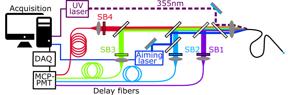
Based on the pulse sampling approach, our group has developed the first multispectral fluorescence lifetime imaging instrument capable of simultaneous measurements of fluorescence lifetime in multiple spectral emission bands suitable for tissue characterization. This instrument consists of a fiber optic probe, a single Microchannel Plate Photomultiplier Tube (MCP-PMT) detector, a fast digitizer and temporal multiplexing scheme using different lengths of fiber optic delay lines for each spectral band. Fast system dynamic range adjustment, real-time data processing and real-time augmentation of fluorescence parameters for intraoperative tissue diagnosis and surgical guidance is achieved. Other groups have adopted the same approach and combined it with galvanometer scanners for both in vitro and in vivo tissue diagnosis with a handheld endoscope or scanning microscopy.
Multi-APDs instrument
To address the limitation of the time-multiplexing scheme, we developed the next generation multi-spectral FLIm instrument utilizing a multi-APDs detection scheme. The new instrument relies on dedicated solid-state UV enhanced silicon avalanche photodiodes (APDs) and gain modulation for each spectral channel allowing channel-wise optimization of detection sensitivity. Fast imaging speed (at least 4 times) is achieved as a result of better quantum efficiency of UV enhanced APD in tissue autofluorescence emission spectral range (400 nm to 700nm). It is also more compact and suitable for clinical use.


Funding
NIH (National Institutes of Health): R01CA187427, R01CA250512